It happens in clinics every week. A therapist has just taken a patient through the core message of pain science — that pain is produced by the nervous system, that it is not a direct readout of tissue damage, and that the body is more robust than it feels. The patient nods along.
Then they ask the question.
"So is my fascia stuck? Should we loosen it up first before I try to move?"
It is a reasonable question, and it exposes a genuine tension inside modern practice. We spend a decade teaching patients that their tissue is not fragile. Then, the moment they feel restricted, both patient and clinician reach for a passive tool to "release" something before movement is allowed.
Cupping sits directly on this fault line. Used one way, it reinforces the idea that something is stuck and must be freed before the body can be trusted. Used another, it opens a short, low-threat window in which a patient is finally willing to move — and the movement is what changes the outcome.
That distinction is the entire argument. The cup is the door. It is not the room.
What Mainstream Practice Already Agrees On
Start with what the guidelines actually say, because they set the terms for everything that follows.
The American Physical Therapy Association's 2021 clinical practice guideline for low back pain is explicit about how pain education and passive techniques are meant to be used together. Pain neuroscience education should be delivered alongside other interventions — exercise or manual therapy — rather than as a stand-alone treatment. Soft tissue mobilization and massage are positioned as short-term tools for pain relief, used in combination with other interventions, not as the intervention itself.
Read that again, because it is not a rejection of passive care. Nothing in the guideline says a passive modality is useless. It says a passive modality has a place — and that the place is subordinate to active care. The evidence-based question is not "does cupping work?" in isolation, but "what does cupping let the patient do that they could not do otherwise?"
Where Fascia Actually Sits
The word "fascia" carries a great deal of marketing baggage, so it is worth being precise about what it is in the literature.
Fascia is a continuous network of connective tissue that wraps muscle, bone, nerve, and organ. It is also densely innervated. Fascial research describes mechanoreceptors — Ruffini endings, Pacinian corpuscles, and interstitial receptors — that respond to sustained stretch, shear, and pressure and relay that information to the central nervous system, where it influences motor tone and body awareness. Some researchers have argued that the fascial network is among the body's richest sensory tissues, with estimates of its nerve supply running into the hundreds of millions of endings.
It also transmits force. Because fascial layers are continuous, load applied in one region is distributed across a much wider network rather than isolated to a single muscle.
Where this becomes clinically interesting is in glide — the ability of adjacent layers to slide on one another. Specialised fibroblasts produce hyaluronic acid, the lubricant that enables that sliding. When the layer becomes denser, gliding is reduced. Ultrasound research has found that people with chronic low back pain had thoracolumbar fascia roughly 20% stiffer than controls, with layers appearing to stick together rather than slide.
Here is the discipline the evidence demands: reduced glide is not the same thing as the source of pain. A stiffened fascial layer is a mechanical and sensory condition. It changes how load is distributed and how the tissue reports itself to the brain. It does not follow that "releasing" it removes pain. For a fuller treatment of this, see our article on fascia as the ignored organ, and the broader science of cupping.
That distinction is what keeps a clinician honest. We are not fixing a stuck structure. We are changing the conditions under which movement becomes possible.
The Door: What Negative Pressure Offers
Most manual therapy compresses tissue. Cupping does the opposite: negative pressure lifts the skin and superficial layers away from the deeper tissue. That single mechanical difference is the whole point.
It matters clinically because of what it does to the patient's threat response. The cup delivers a strong, novel, but controllable sensory input — and it does so without the pain that a rigid instrument bearing down on a sensitive region would provoke. For a patient who is guarding, that is a window.
The technique also has a mainstream lineage that is frequently overlooked. Myofascial decompression (MFD) — cupping applied over areas of assessed restriction and combined with active movement — was developed within American sports physical therapy by Christopher DaPrato at UC Berkeley and UC San Francisco. It is taught in UCSF's orthopedic residency program and was used to prepare Olympic medical staff before the 2016 Rio Games. This is not an alternative technique imported from outside the profession; it is one the profession built. We cover the movement-based rationale in more depth in Functional Cupping and Neuromuscular Re-education.
The movement-based evidence is early, but it is not empty. A 2020 pilot randomised controlled trial in the International Journal of Sports Physical Therapy compared MFD with foam rolling in collegiate athletes with hamstring pathology. Both approaches improved range of motion acutely; patients rated MFD more favourably on global perceived change. That is a small, single-session study with short follow-up, and it should be read as such. But it is consistent with the mechanism: the tool creates the condition for movement, and the movement does the work.
The Evidence, Stated Honestly
If this argument is going to survive scrutiny, it has to face the strongest evidence against it, not just the evidence in its favour.
Cupping for chronic musculoskeletal pain. A 2025 systematic review and meta-analysis in BMJ Open pooled randomised trials and found a significant effect on pain intensity (standardised mean difference −1.17; 95% CI −1.93 to −0.42), graded as moderate-certainty evidence. It found no significant improvement in functional disability (SMD −0.24; 95% CI −0.93 to 0.46; p = 0.51) and no significant improvement in mental health (SMD 0.08; 95% CI −0.12 to 0.27; p = 0.46). The analysis was limited to immediate effects, heterogeneity was high (I² = 94%), and it included only ten studies.
Read that carefully. Cupping reliably moves pain intensity in the short term. On its own, it does not restore function.
Cupping versus sham. An earlier meta-analysis in The Journal of Pain found a large short-term effect of cupping against no treatment (SMD −1.03) — but no significant effect against sham cupping (SMD −0.27; 95% CI −0.58 to 0.05). This is the honest ceiling of the evidence base. A convincing sham for suction is genuinely difficult to construct, so the result is not proof of pure placebo. But it does mean we cannot claim the effect is independent of expectation and attention.
Pain education plus exercise. Set against that, the intervention that does accumulate supporting evidence is education combined with active care. A 2022 meta-analysis in Pain found that pain neuroscience education plus exercise outperformed exercise alone for short-term pain, disability, kinesiophobia, and catastrophizing — though certainty ranged from moderate (kinesiophobia) to low or very low (pain, disability, catastrophizing).
Put the two evidence lines side by side and the roles become obvious. Cupping can lower pain intensity now. Movement is what changes function later. Neither finding is a reason to abandon the other. They describe a sequence.
Making the Doorway Operational
If a cup is a doorway, the clinical task is to walk the patient through it. In practice that means a sequence rather than a technique:
- Assess first. Identify the specific movement or region that is restricted or threatening. Not a fixed point, not a blanket application.
- Dose the decompression short. A few minutes of low-to-moderate suction is enough to open the window. More is not better; longer holds and higher pressures mainly increase the marks and the discomfort.
- Move inside the window. With cups in place or immediately after, take the patient through the movement they had been avoiding — small range, low load, tolerable.
- Progress the load. The doorway is temporary by design. Strength and graded exposure carry the adaptation.
- Send it home. A short self-directed programme, with or without a cup, so that the patient owns the change.
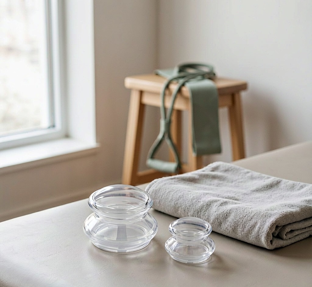
Tool design is not incidental to this model. Three features make dose-and-move genuinely executable in a clinic: controllable suction, so pressure is tuned to the patient's tolerance rather than to the pump's maximum; soft silicone that deforms and glides, so movement can continue while the cup stays sealed; and transparent cups, which let the clinician observe the skin's response during the session rather than after it. These are the properties we build into the Classic Mushroom and Clear Straight series, because they are what the clinical sequence actually requires.
The Language Problem
There is one further requirement, and it is the part clinics most often get wrong: the script.
If your pain education tells a patient that pain is not a measure of damage, then a cupping script that treats marks as a measure of something removed is a direct contradiction. The two messages cannot coexist in the same treatment room. A patient who has just been taught to stop scanning their body for signs of harm will read a dark mark as evidence of harm — unless the clinician pre-empts it.
The accurate description is straightforward. The marks are a local capillary response to negative pressure: petechiae and mild ecchymosis — the same mechanism as a bruise, but produced by a pulling force rather than an impact. They are expected, they are not a sign of tissue damage, and they typically fade within several days. They are not a measure of anything that was "released," "drawn out," or "cleared." For patient-facing wording, see What Cupping Marks Actually Mean and our breakdown of the lymphatic detox myth.
That single adjustment — describing what the mark is, rather than what it supposedly proves — is what keeps the tool consistent with the education sitting right beside it.
Where This Leaves the Clinician
Cupping is not pain education, and it is not a structural fix. It is a short-term, low-threat way to make movement possible — and movement, combined with education, is where durable change comes from.
The cup opens the door. The therapy happens on the other side.
Equip the Doorway, Not the Bruise
ELERA supplies medical-grade silicone cupping systems designed for clinical dosing — controllable suction, soft gliding edges, and transparent cups for real-time observation. Contact our B2B team for wholesale pricing, clinical bulk orders, and OEM customization.
Partner With UsReferences
- George SZ, Fritz JM, Silfies SP, et al. Interventions for the Management of Acute and Chronic Low Back Pain: Revision 2021. J Orthop Sports Phys Ther. 2021;51(11):CPG1–CPG60. doi:10.2519/jospt.2021.0304.
- Jia Y, Dong X, Chai Y, Bai Z, Sun T, Hou X. Effects of cupping therapy on chronic musculoskeletal pain and collateral problems: a systematic review and meta-analysis. BMJ Open. 2025;15(5):e087340. doi:10.1136/bmjopen-2024-087340.
- Cramer H, Klose P, Teut M, et al. Cupping for Patients With Chronic Pain: A Systematic Review and Meta-Analysis. J Pain. 2020;21(9–10):943–956. doi:10.1016/j.jpain.2020.01.002.
- Siddall B, Ram A, Jones MD, Booth J, Perriman D, Summers SJ. Short-term impact of combining pain neuroscience education with exercise for chronic musculoskeletal pain: a systematic review and meta-analysis. Pain. 2022;163(1):e20–e30. doi:10.1097/j.pain.0000000000002308.
- Warren AJ, LaCross Z, Volberding JL, O'Brien MS. Acute outcomes of myofascial decompression (cupping therapy) compared to self-myofascial release on hamstring pathology after a single treatment. Int J Sports Phys Ther. 2020;15(4):579–592.
- Langevin HM, Stevens-Tuttle D, Fox JR, et al. Ultrasound evidence of altered lumbar connective tissue structure in human subjects with chronic low back pain. BMC Musculoskelet Disord. 2009;10:151. doi:10.1186/1471-2474-10-151.
- Schleip R. Fascial plasticity — a new neurobiological explanation: Part 1. J Bodyw Mov Ther. 2003;7(1):11–19. doi:10.1016/S1360-8592(02)00067-0.